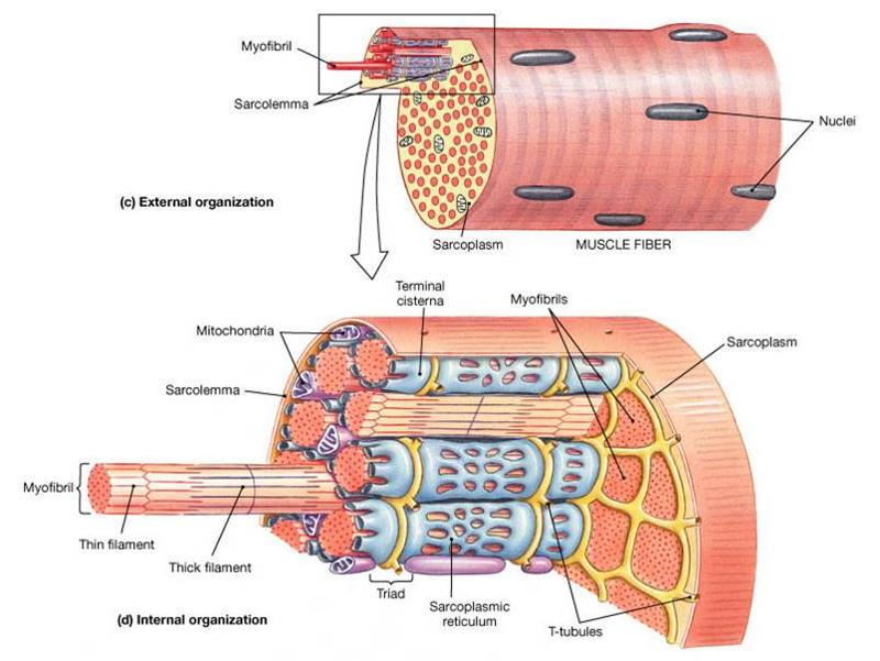
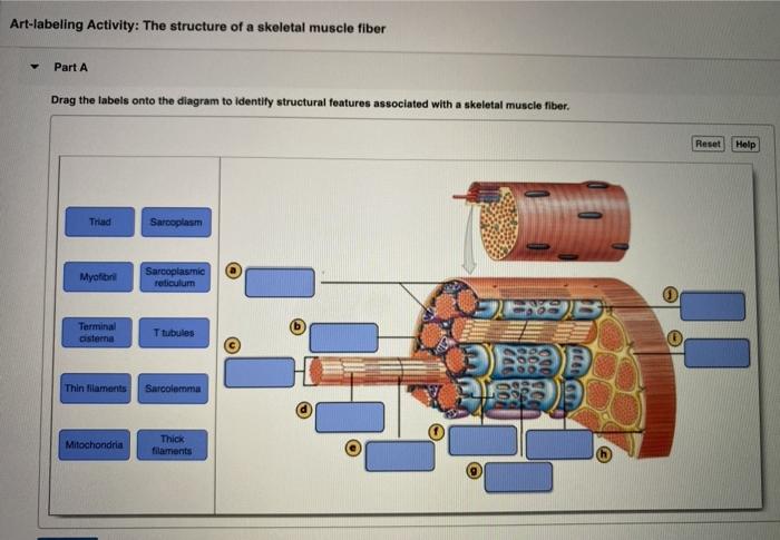
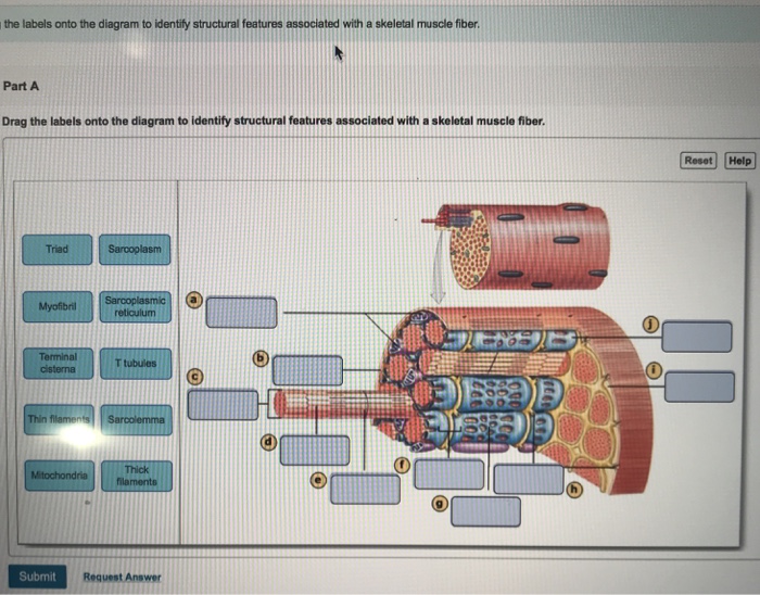
42 art-labeling activity: the structure of a skeletal muscle fiber
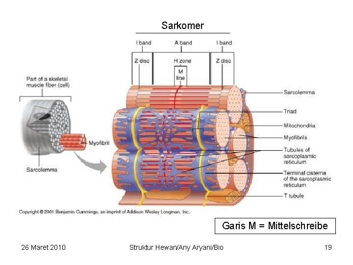
A & P Ch 6 Musclular System Student PPT - SlideShare Microscopic Anatomy of Skeletal Muscle 4. Describe the microscopic structure of skeletal muscle and explain the role of actin- and myosin- containing myofilaments and label a sarcomere diagram. Skeletal Muscle Activity 5. Describe how an action potential is initiated in a muscle cell. (neuromuscular junction, acetylcholine, Ca++…) 6. (Get Answer) - Art-labeling Activity: Figure 27.11b. Art-labeling ... Art-Labeling Activity: The Structure Of A Sarcomere Part A Drag The Labels To The Appropriate Location In The Figure. Reset Help A Band Barmere Hand Band MI Art-Labeling Activity: The Structure Of A Skeletal Muscle Fiber Part A Drag The Labels Onto...
Art-labeling Activity: The Structure of a Skeletal Muscle Fiber Start studying Art-labeling Activity: The Structure of a Skeletal Muscle Fiber. Learn vocabulary, terms, and more with flashcards, games, and other study tools. Search. Create. ... The Structure of a Skeletal Muscle Fiber... OTHER SETS BY THIS CREATOR. Pathophysiology. 11 terms. BabeRuthless0504. Lympathetic System. 37 terms.

Art-labeling activity: the structure of a skeletal muscle fiber
Bsc2085l chapter 013 activity 1 skeletal muscle - Course Hero BSC2085L Chapter 013 Activity 1 Skeletal Muscle Organization-005 Part A The area of a sarcomere where the thin actin filaments connect to one another is called the _____. ANSWER: Correct The Z line or Z disc consists of proteins called actinin that anchor the actin filaments together. A message from your instructor... Activity 2: The Neuromuscular Junction Art-labeling Activity: Skeletal ... chapter 9 Flashcards | Quizlet Art-labeling Activity: The structure of a skeletal muscle fiber PICTURE Chapter Test - Chapter 9 Question 3 Which thin-filament-associated structure is distinguished by its constituents of three globular subunits, one of which has a receptor that binds two calcium ions? a) G-actin b) nebulin c) tropomyosin d) troponin D ... (Get Answer) - Art-labeling Activity: Figure 19.2b (2 of 2) Drag the ... Art-labeling Activity: Figure 19.2b (2 of 2) Drag the appropriate labels to their respective targets. Reset Help Collagen fibers Artery Lumen Basement membrane Endothelial cells Smooth muscle and elastic fiber Capilary network Internal elastic membrane Endothelium External lasti membrane Capillary Vasa vasorum Subendothelal layer.
Art-labeling activity: the structure of a skeletal muscle fiber. (Get Answer) - Art-labeling Activity:. Art-labeling Activity: | Transtutors Art-Labeling Activity: The Structure Of A Sarcomere Part A Drag The Labels To The Appropriate Location In The Figure. Reset Help A Band Barmere Hand Band MI Art-Labeling Activity: The Structure Of A Skeletal Muscle Fiber Part A Drag The Labels Onto... PDF The Muscular System Tour Lab The Muscular System - lcboe.net is broken down to provide energy. To help delay muscle fatigue, the muscle fibers are constantly switching on an off to allow individual fibers a moment to rest. This activity will demonstrate the effects of action of muscle fibers. Do this: 1. Hold a popsicle stick in front of you , parallel to the table top. 2. Place a bent paper clip on the ... A&P 1- CHAPTER 9 MASTERING ASSIGNMENTS Flashcards - Quizlet Art-labeling Activity: The structure of a skeletal muscle fiber PICTURE Which thin filament-associated protein binds two calcium ions? troponin Action potential propagation in a skeletal muscle fiber ceases when acetylcholine is removed from the synaptic cleft. Solved Art-labeling Activity: The Structure of Skeletal and - Chegg See the answer Art-labeling Activity: The Structure of Skeletal and Cardiac Muscle Fibers Drag the labels to the appropriate location in the figure. Show transcribed image text Expert Answer 100% (2 ratings) The first picture is of a skeletal muscle which can identified by parallel bun … View the full answer
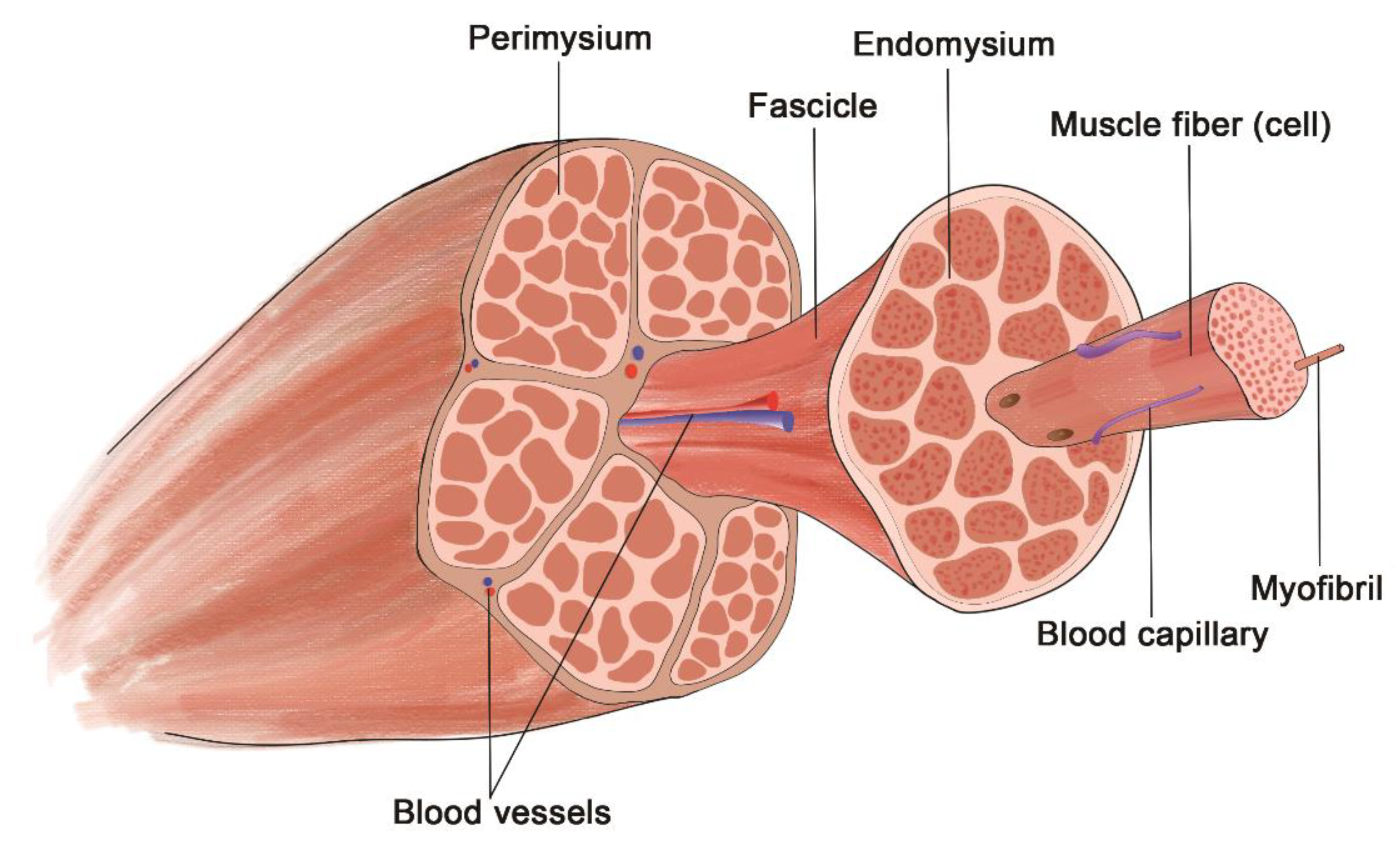
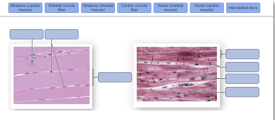
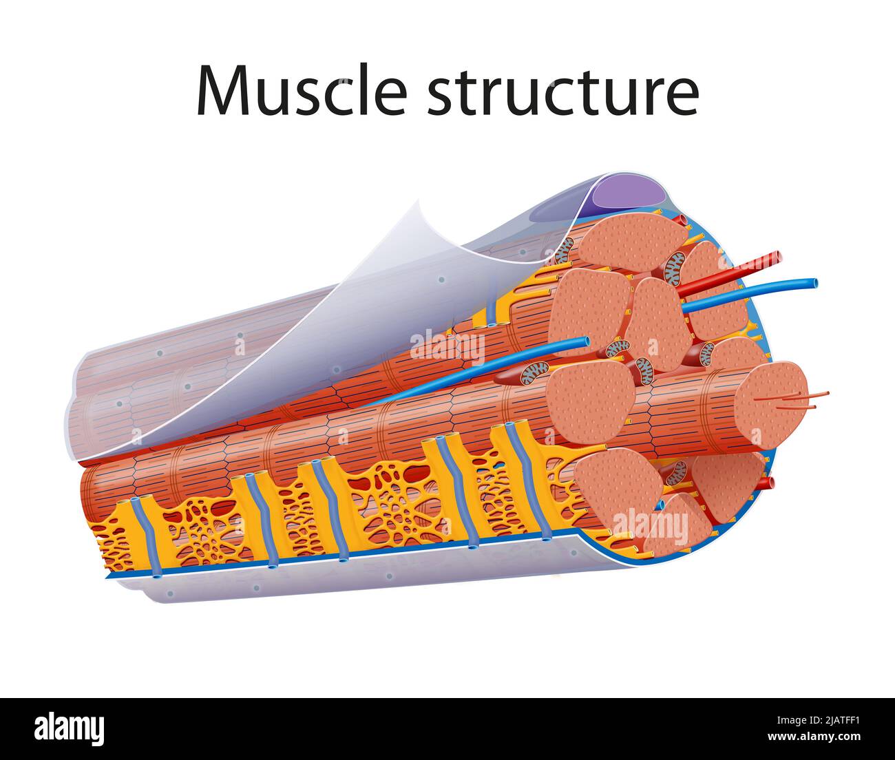
Bio 2331 Prelab 6 Muscles Part 1.pdf - 2/10/22, 10:55 PM... ANSWER:skeletal muscle cells consist of cylindrical, non-branched cells with multiple nuclei skeletal muscle cells possess cell junctions called intercalated discs skeletal muscle cells are branched and generally possess one nucleus skeletal muscle cells are spindle-shaped and possess one centrally located nucleus PDF In this chapter, you will learn that - Pearson 9.2 A skeletal muscle is made up of muscle fibers, nerves, blood vessels, and connective tissues Learning Objective Describe the gross structure of a skeletal muscle. For easy reference, Table 9.1 on p. 286 summarizes the levels of skeletal muscle organi-zation, gross to microscopic, that we describe in this and the following modules. Answer correct art based question chapter 4 question - Course Hero ANSWER: Correctmultinucleate cells branched cells intercalated discs situated between cells striations tendons and ligaments attached to bones heart ducts of certain glands dense irregular connective tissue smooth muscle tissue skeletal muscle tissue cardiac muscle tissue BIO 200 Chapter 9 - Muscle Tissue Physiology Flashcards - Quizlet The storage and release of calcium ions is the key function of the: sarcoplasmic reticulum. A group of skeletal muscle fibers together with the surrounding perimysium form a (n): fascicle. Art-Ranking Activity: Stages of an action potential. A crossbridge forms when: a myosin head binds to actin.
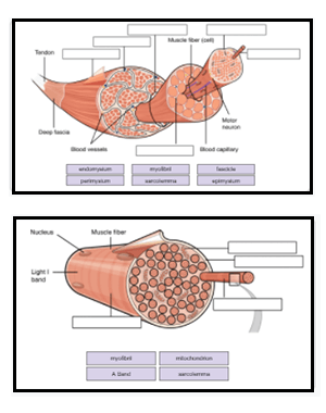
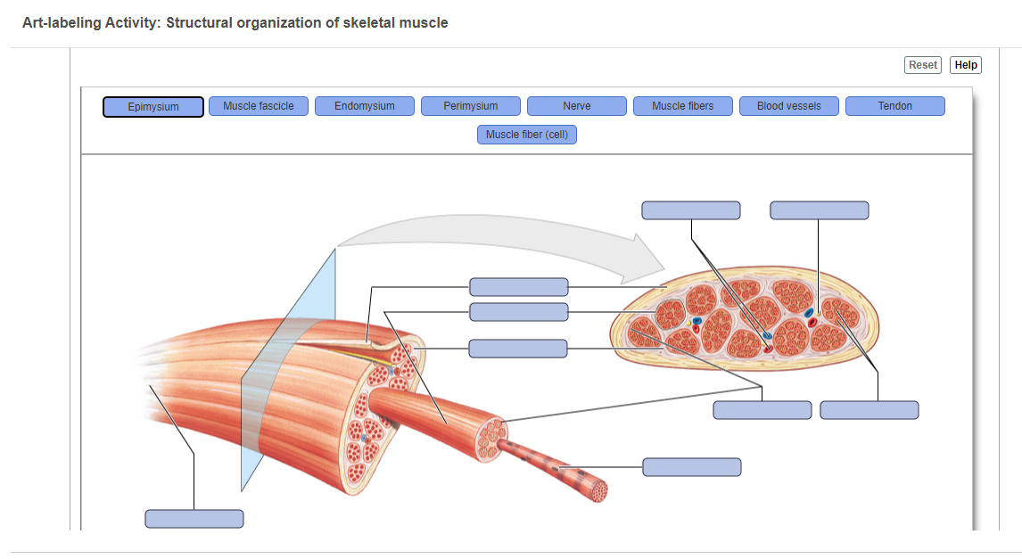
Answered: Art-labeling Activity: Structural… | bartleby Answered: Art-labeling Activity: Structural… | bartleby. Homework help starts here! Science Biology Q&A Library Art-labeling Activity: Structural organization of skeletal muscle Reset Epimysium Muscle fascicle Endomysium Perimysium Nerve Muscle fibers Blood vessels Tendon Muscle fiber (cell) Week 3 Chapter 9.pdf - 4/23/22, 5:03 PM Week 3 Chapter 9... The tension produced by a contracting skeletal muscle fiber results from the interaction between the thick and thin filaments within sarcomeres. The mechanism of skeletal muscle contraction is explained by the sliding filament theory Read through Spotlight Figure 9.7, and then complete the questions and activity below. Part A - Initiation of Contraction Contraction is initiated by release of ... Art-Labeling Activity: The Structure Of A Sarcomere Part A Drag The ... Reset Help A Band Barmere Hand Band MI Art-Labeling Activity: The Structure Of A Skeletal Muscle Fiber Part A Drag The Labels Onto The Diagram To Identity Structural Features Associated With A Skeletal Muscle Fiber. Reset Help Trad Apr 01 2022 08:57 AM Expert's Answer Solution.pdf Next Previous Q: Q: Q: Q: chapter 9 Flashcards | Quizlet Art-labeling Activity: The structure of a skeletal muscle fiber PICTURE Chapter Test - Chapter 9 Question 3 Which thin-filament-associated structure is distinguished by its constituents of three globular subunits, one of which has a receptor that binds two calcium ions? a) G-actin b) nebulin c) tropomyosin d) troponin D ...
A&P 1- CHAPTER 9 MASTERING ASSIGNMENTS Flashcards | Quizlet
(Solved) - Art-Labeling Activity: Functions of antibodies ... - Transtutors Art-Labeling Activity: ...
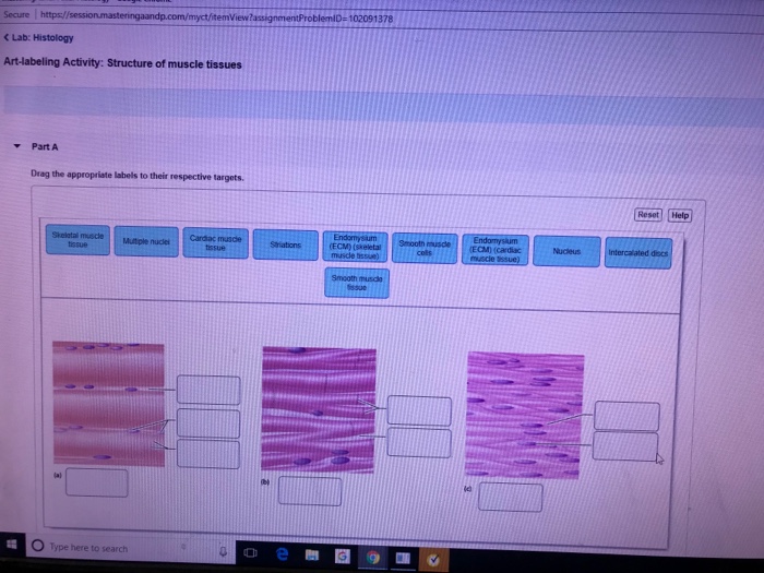
Solved Secure https:/ C Lab: Histology Art-labeling | Chegg.com
art-labeling activity: the structure of the digestive tract An unregistered player played the game 29 seconds ago. 2018-7-14 Art-labeling Activities Use the art-labeling activities to quiz yourself on key anatomical structures in this chapter. Structural organization of skeletal muscle Reset Help Epimysium Muscle fascicle Endomysium Perimysium Nerve Muscle fibers Blood vessels Tendon Muscle fiber cell.
Muscles Labeling
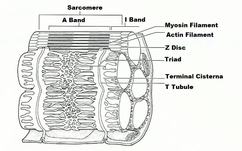
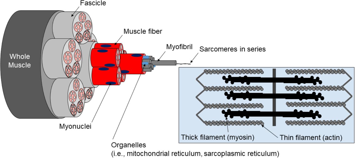
Skeletal Muscle Fiber Structure and Function - Open Textbooks for Hong Kong The striated appearance of skeletal muscle tissue is a result of repeating bands of the proteins actin and myosin that occur along the length of myofibrils. Myofibrils are composed of smaller structures called myofilaments. There are two main types of myofilaments: thick filaments and thin filaments.
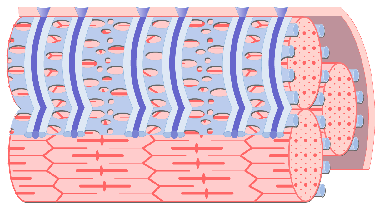
Internal Anatomy of Skeletal Muscle Fibers | GetBodySmart
Solved Art-labeling activity: structure of skeletal muscle - Chegg This problem has been solved! See the answer. See the answer See the answer done loading. Art-labeling activity: structure of skeletal muscle fiber. Drag the appropriate lablels to their respective targets. Expert Answer.

The Muscular System. - ppt download
10.2 Skeletal Muscle - Anatomy and Physiology 2e | OpenStax These tissues include the skeletal muscle fibers, blood vessels, nerve fibers, and connective tissue. Each skeletal muscle has three layers of connective tissue (called "mysia") that enclose it and provide structure to the muscle as a whole, and also compartmentalize the muscle fibers within the muscle ( Figure 10.3 ).
Muscles and Muscle Tissue
BIOL.docx - Ch9 Hmwk Art-labeling Activity: Structural ... - Course Hero View Notes - BIOL.docx from BIOL 2533 at Fayetteville State University. Ch9 Hmwk Art-labeling Activity: Structural organization of skeletal muscle previous 3 of 8 next You completed this

Ch 10 lab map Flashcards | Quizlet
(Get Answer) - Art-labeling Activity: Figure 19.2b (2 of 2) Drag the ... Art-labeling Activity: Figure 19.2b (2 of 2) Drag the appropriate labels to their respective targets. Reset Help Collagen fibers Artery Lumen Basement membrane Endothelial cells Smooth muscle and elastic fiber Capilary network Internal elastic membrane Endothelium External lasti membrane Capillary Vasa vasorum Subendothelal layer.
OVERVIEW OF MUSCLE TISSUE
chapter 9 Flashcards | Quizlet Art-labeling Activity: The structure of a skeletal muscle fiber PICTURE Chapter Test - Chapter 9 Question 3 Which thin-filament-associated structure is distinguished by its constituents of three globular subunits, one of which has a receptor that binds two calcium ions? a) G-actin b) nebulin c) tropomyosin d) troponin D ...
Muscles and Muscle Tissue
Bsc2085l chapter 013 activity 1 skeletal muscle - Course Hero BSC2085L Chapter 013 Activity 1 Skeletal Muscle Organization-005 Part A The area of a sarcomere where the thin actin filaments connect to one another is called the _____. ANSWER: Correct The Z line or Z disc consists of proteins called actinin that anchor the actin filaments together. A message from your instructor... Activity 2: The Neuromuscular Junction Art-labeling Activity: Skeletal ...

Skeletal Muscle: Structure and Contraction | BIO103: Human ...
Exercise 14: Microscopic Anatomy and Organization of Skeletal ...
Anatomy Exam 2 Flashcards - Easy Notecards

A&P 1- CHAPTER 9 MASTERING ASSIGNMENTS Flashcards | Quizlet
Solved
Muscle Tissue. - ppt download

PDF) ITAP: Clinical outcomes and implant design optimisation ...
Solved -ling Activity: Structure of a Skeletal Muscle Fiber ...

BIO 200 Chapter 9 - Muscle Tissue Physiology Flashcards | Quizlet

Art labeling Activity: Figure 19.1b (1 of 2) - PDF Free Download
Differential response of oxidative and glycolytic skeletal ...
Solved Art-labeling Activity: The Structure of a Sarcomere ...

PDF) Striated muscle function, regeneration, and repair
Polymers | Free Full-Text | A Review of Recent Advances in ...

Mastering A&P II Chapter 25 - The Urinary System Diagram ...
Solved Art-labeling Activity: The Structure of Skeletal and ...
Answered: Art-labeling Activity: Structural… | bartleby

final Flashcards | Quizlet
Skeletal muscle hi-res stock photography and images - Alamy
26 Maret 2010 Struktur HewanAny AryaniBio 1 Jaringan

6 The Muscular System. - ppt video online download

Basic anatomy and physiology, Medical anatomy, Human bones ...
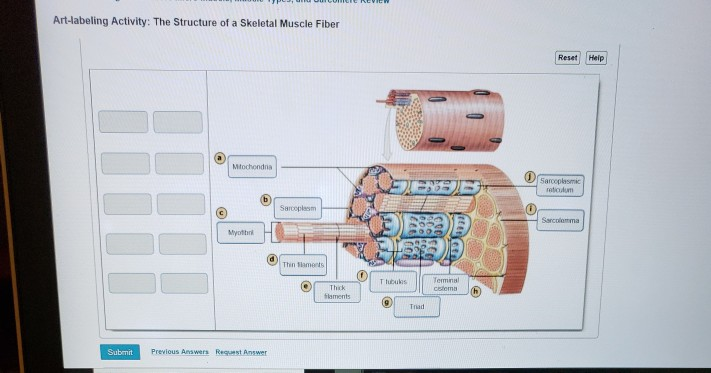
Solved Art-labeling Activity: The Structure of a Skeletal ...

Final Exam Set Flashcards | Quizlet
Frontiers | A Critical Evaluation of the Biological Construct ...
Support Systems | Biology for Majors II | | Course Hero

Ex 12 Microscopic Anatomy & Organization of Skeletal Muscle ...
The Muscular System The Muscular System

Here we see a myogram displaying a muscle twitch. A typical ...

A&P 1 exam 2: muscle tissue Diagram | Quizlet
Solved
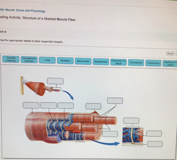
Solved Art-labeling activity: structure of skeletal muscle ...
Solved the labels onto the diagram to identify structural ...
Post a Comment for "42 art-labeling activity: the structure of a skeletal muscle fiber"