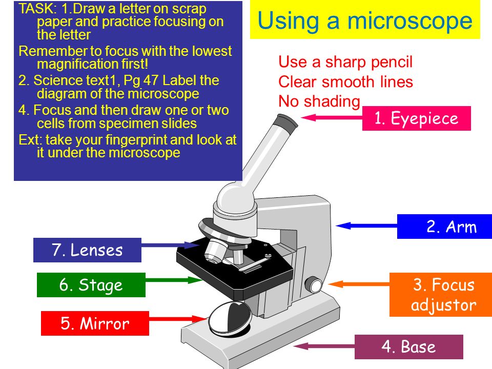
44 draw and label microscope
Microbiology Virtual Lab I - Amrita Vishwa Vidyapeetham The medium mainly used for this purpose is SIM medium ( Sulphide Indole Motility medium) which is a combination differential medium that tests three different parameters, Sulfur Reduction, Indole Production and Motility. This media has a very soft consistency that allows motile bacteria to migrate readily through them causing cloudiness. Microscope Quizlet Use And Worksheet Parts to use a microscope, pick up a prepared slide by its edges and place it on the microscope's stage worksheet parts of a microscope use the fine adjustment knob to bring the specimen into focus use the page up (pgup) and page down (pgdn) keys to get used to scrolling in a worksheet use a different type of microscope use a different type of …
Mitosis Bead Lab Mitosis in an Onion Root - view slides and count the number of cells visible in each stage Mitosis was observed and timed in Lab 3A I had trouble finding brown beads, which I would have preferred to use for the eye genes (instead of blue and grey) Mitosis is nuclear division plus cytokinesis, and Lab 9: Mitosis and Meiosis - Biology LibreTexts Lab 9: Mitosis and Meiosis - Biology LibreTexts.

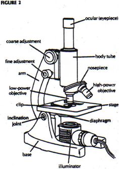
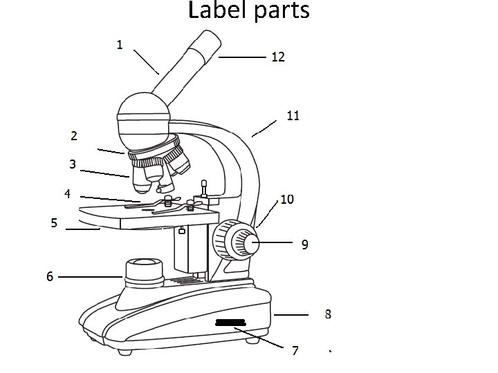
Draw and label microscope
Encouragement: Be Still "Be still in the presence of the LORD, and wait patiently for him to act." (Psalms 37:7a NLT) Sorry about the poor image. I should have ta... DeepBacs for multi-task bacterial image analysis using open-source deep ... DeepBacs guides users without expertise in machine learning methods to leverage state-of-the-art artificial neural networks to analyse bacterial microscopy images. The best Prime Day deals for teachers, starting at $6 Sharpens colored pencils and No. 2s alike in seconds flat. Leave it to blade-master X-Acto to make a fierce pencil sharpener. And it's got 6,600 five-star ratings. One very impressed pleased...
Draw and label microscope. Cerebral cortex: Structure and functions | Kenhub The mesocortex is a transitional form between the allocortex and isocortex. It contains three to six layers and is found in the insula, cingulate and parahippocampal gyri. The neocortex, as its name suggests, is the most recent cortical region and makes up to 90% of the human cortex. Biology Chapter 12 Section 3 microscope), (b) onion cells (viewed with a light microscope), and (c) Vibrio tasmaniensis bacterial cells (viewed using a scanning electron microscope) are from very different organisms, yet all share certain characteristics of basic cell structure. (credit a: modification of work by Ed Uthman, MD; credit b: modification of work by Umberto ECLIPSE Ti2 Series | Inverted Microscopes | Products | Nikon ... The ECLIPSE Ti2 inverted microscope delivers an unparalleled 25mm field of view (FOV) that revolutionizes the way you see. With this incredible FOV, the Ti2 maximizes the sensor area of large-format CMOS cameras without making compromises, and significantly improves data throughput. Onion Structure Cell Search: Onion Cell Structure. Some of the cell walls are very thin in some plants 393 362 123 Cell Biology Study Guide: pg The last part of the lesson requires pupils to follow the instructions included in the PowerPoint to prepare an onion slide to then observe plant cells under the microscope down the circuit in fixed-size cells, which are unwrapped by a symmetric key at each node (like the ...
Spinal cord: Anatomy, structure, tracts and function | Kenhub It shows four surfaces: anterior, posterior, and two lateral. They feature fissures (anterior) and sulci (anterolateral, posterolateral, and posterior). The gray matter is the butterfly-shaped central part of the spinal cord and is comprised of neuronal cell bodies. It shows anterior, lateral, and posterior horns. Diatom - Wikipedia Diatom (Neo-Latin diatoma) refers to any member of a large group comprising several genera of algae, specifically microalgae, found in the oceans, waterways and soils of the world.Living diatoms make up a significant portion of the Earth's biomass: they generate about 20 to 50 percent of the oxygen produced on the planet each year, take in over 6.7 billion metric tons of silicon each year from ... Of Function Microscope Quiz And Parts 1) please label the parts of the microscope below by putting the letter that matches the location on the microscope parts of a microscope - in this topic, we will now know and identify the parts of a microscope and discuss each of their functions the stage and its function is to hold the glass slide keyword research: people who searched parts of … Worksheet Use Quizlet And Microscope Parts to use a microscope, pick up a prepared slide by its edges and place it on the microscope's stage in addition, we needed to look at contrasts of some specimens in this lab you are not authorised to view the member list or profiles foot or base microscope quiz pdf use the word list to help you label the microscope microscope quiz pdf use the word …
Optically Manipulated Neutrophils as Native Microcrafts In Vivo Figure 1. Figure 1. (a) Schematic illustration for optically manipulated neutrophil microcraft in vivo. (b) Optical microscopic images of zebrafish (b1) and fluorescence-labeled neutrophils (b2 and b3). (c) Dynamic manipulation of resting neutrophils (c1-c4) with a detailed velocity (c5) and acquired maximum motion velocity for shifting the ... Shimadzu Corporation July 12, 2022 Using AI to Support Working Practice Reforms at Research Sites Release of Peakintelligence for GCMS: AI Software for GC-MS/MS Product & Event info News & Notices; July 12, 2022 Release of a New Type of Mobile X-ray System for Outside of Japan Accommodating a Wide Range of Needs for Mobile X-ray Systems Product & Event info News & Notices; July 1, 2022 Detecting Flaws within ... Localizing Proteins on Single Trafficking Organelles in 3D with ... This labeling method allows for the global nanoscale marking of proteins in and around organelles in 3D inside cells. 2 Materials 2.1 Cell Culture and Transfection 1. Cells of interest: HeLa (American Type Culture Collection), and PC12-GR5 cells line—originated from R. Nishi, P. Stork, and W. Almers (OHSU, Portland, Oregon). 2. scheme work biology - Free KCPE Past Papers Introduction to light microscope. By the end of the lesson, the learner should be able to: Define a cell; Draw and label the light microscope; Description of a cell; Drawing and labeling the light microscope . Light microscope; Diagram of light microscope; Comprehensive secondary Biology students Bk. 1 page 17; Teachers bk. 1 pages 11-19; KLB ...
Microscope - diagram Tom Butler | Science skills, Microscope ...
And Worksheet Microscope Parts Use Quizlet to use a microscope, pick up a prepared slide by its edges and place it on the microscope's stage includes capitalizing the first word of sentences, proper nouns, place names, days and holidays and the titles of books, movies and brand names when you are finished using the microscope: a total magnification = 2 in this biology lesson, students …
Label Parts Of Microscope - ClipArt Best
Light Microscope (Theory) - Amrita Vishwa Vidyapeetham Carry the microscope by holding the C-shaped arm with one hand and other hand under the base. Never swing the microscope while carrying. Never allow direct light to fall on the microscope. Cover the microscope with a plastic cover when not in use. While using oil immersion objective, do not adjust the coarse screw.
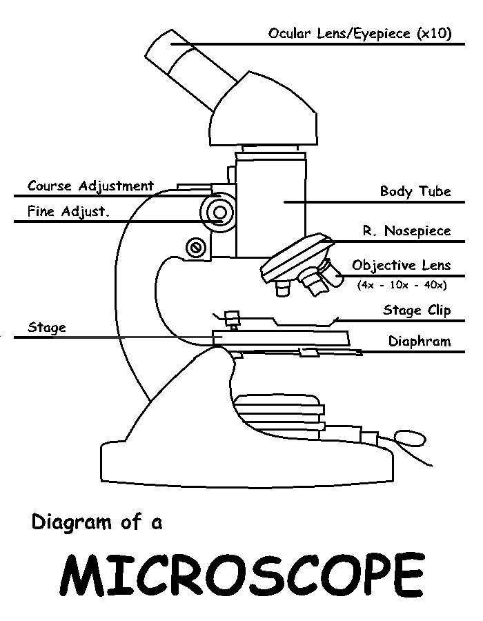
Diagram of a Microscope by ScienceDoodles on DeviantArt
Aqua hold pap pen - ProSciTech Aqua hold pap pen. Antigen tests. Liquid-repellent slide marker pens for staining procedures; 5mm pen tip assures broad band to retain fluids. The liquid blocker pen is very resistant to PBS, TBS, and TBST in immuno-staining procedures. Draw easy to see red hydrophobic wells on slides. Once dry, the barrier is insoluble in alcohol or acetone.

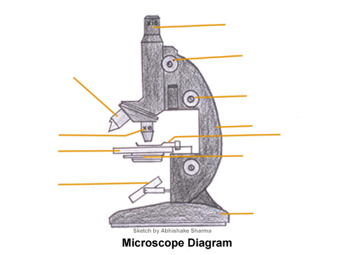
How to draw the diagram of compound microscope - Brainly.in
Parts Microscope Function And Quiz Of microscope to label some of the worksheets for this concept are label parts of the microscope answers the microscope parts and use label parts of the microscope parts of the light microscope labeling scientific tools microscope name use the word list to help you label the 12 7 3 science light microscope wo rk name period date lab 3 use of the …
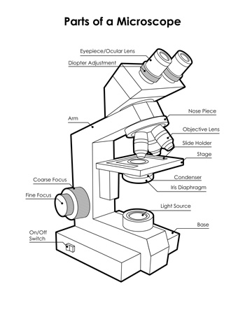
Microscope, Microscope Parts, Labeled Diagram, and Functions
And Use Worksheet Parts Microscope Quizlet cut out the letter "e" and place it on the slide face up com when the microscope uses glass slides, it will first take a thin, transparent portion of glass as its slide and then move it over a microscope slide holder that is attached to the slide holder double 2x10 floor joist span looking for the right diagramming sentences worksheet to engage …

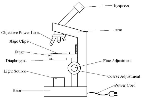
Label Microscope Diagram - EnchantedLearning.com
And Of Parts Microscope Function Quiz name the part of the microscope that you would place the slide on for viewing microscope parts and functions draft students will observe onion cells under a microscope 1) please label the parts of the microscope below by putting the letter that matches the location on the microscope components of this lesson take students through names of …

Simple Microscope - Diagram (Parts labelled), Principle ...
5 White Blood Cells Types and Their Functions - New Health Advisor There are two different kinds of white blood cells and each looks different from one another under the microscope. These include granulocytes and agranulocytes. Granulocytes have visible granules or grains inside the cells that have different cell functions. Types of granulocytes include basophils, neutrophils, and eosinophils.

Directions: Draw and label the parts of the microscope.(Name ...
Visual recognition of social signals by a tectothalamic neural circuit Fast volumetric imaging of the tectum and/or thalamus was performed using a custom-built remote focusing arm added before the microscope (Extended Data Fig. 3b). The remote focusing path was ...

Parts of a Microscope with Their Functions • Microbe Online
5 Steps of Gram Staining Procedure: How to Interpret the Results 1. Prepare the Glass Microscopic Slide To make sure that your slides are grease/oil-free, you need to first wash them with soap and water then wipe slides with alcohol. This will remove any fingerprints or dirt from them. Dry the slides and put them on lab towels until they are ready to be used. 2. Label the Slides
Free Microscope Drawing, Download Free Microscope Drawing png ...
'God Bless America ETF' May Be Coming Soon With the Ticker YALL BC-'God-Bless-America-ETF'-May-Be-Coming-Soon-With-the-Ticker-YALL , Katie Greifeld. (Bloomberg) -- An exchange-traded fund is in the works that is looking at brandishing the made-in-the-USA label. But it's an American fund with a twist -- it will remove any companies the firm deems "activist.". The God Bless America ETF, advised by ...

Easy labeled diagram of Microscope - YouTube
The best Prime Day deals for teachers, starting at $6 Sharpens colored pencils and No. 2s alike in seconds flat. Leave it to blade-master X-Acto to make a fierce pencil sharpener. And it's got 6,600 five-star ratings. One very impressed pleased...

Parts of Microscope
DeepBacs for multi-task bacterial image analysis using open-source deep ... DeepBacs guides users without expertise in machine learning methods to leverage state-of-the-art artificial neural networks to analyse bacterial microscopy images.
![Very Easy!!! How to draw microscope [with labels] - YouTube](https://i.ytimg.com/vi/mtXYI7wO-qs/maxresdefault.jpg)
Very Easy!!! How to draw microscope [with labels] - YouTube
Encouragement: Be Still "Be still in the presence of the LORD, and wait patiently for him to act." (Psalms 37:7a NLT) Sorry about the poor image. I should have ta...

Google Image Result for https://rsscience.com/wp-content ...
Cells and Microscopes LO:- use and label a microscope - draw ...

how to draw microscope step by step slow and medium speed
Microscope With Labels clip art Vectors graphic art designs ...

Course: s4: Biology , Topic: UNIT 3: MICROSCOPY

label microscope diagram | Charts | Microscope, Anatomy bones ...

Microscope Drawing | How To Draw A Microscope Diagram | Easy And Simple Step By Step Tutorial

How to Draw a Microscope and Label Its Parts

How to draw Microscope diagram for beginners - step by step

Microscope Drawing Worksheet | Clipart library - Free Clipart ...

Parts Of A Microscope Drawing Grade - Gr 9 Natural Science ...

How to draw a microscope step by step | Drawings, Easy ...

Compound Microscope Parts – Labeled Diagram and their ...
Free Microscope Drawing, Download Free Microscope Drawing png ...

Draw a labelled diagram of a compound microscope.
Free Microscope Drawing, Download Free Microscope Drawing png ...
Microscope. Vector drawing stock vector. Illustration of ...

Microscope Diagram Diagram | Quizlet

Living Environment Course

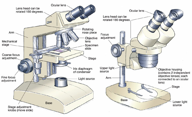
Types of Microscopes: Definition, Working Principle, Diagram ...

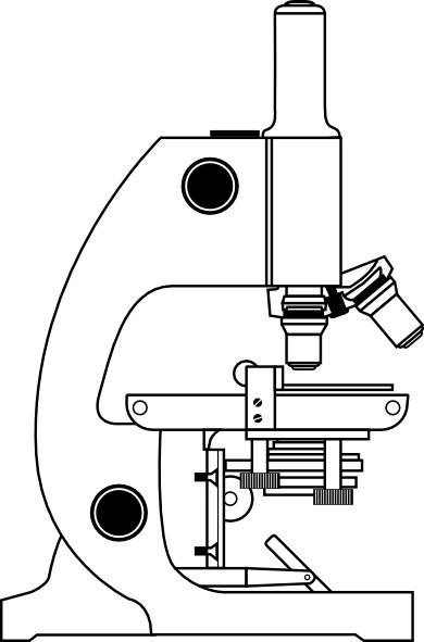
Unlabeled Microscope Diagram posted by Christopher Thompson
Compound Microscope Drawing - ClipArt Best - ClipArt Best ...
Compound Microscope Review Label parts 1 body tube
Free Microscope Drawing, Download Free Microscope Drawing png ...

BIOLOGY FROM 1 | EQUIPMENTS USED FOR OBSERVATION | Cours ...

Microscope With Labels Clip Art at Clker.com - vector clip ...
Microscope Diagram Labeled, Unlabeled and Blank | Parts of a ...

Microscope Labeling

Microscope Diagram - Label Diagram | Quizlet

microscope vector sketch 7312430 Vector Art at Vecteezy

Simple Microscope - Diagram (Parts labelled), Principle ...
Post a Comment for "44 draw and label microscope"