38 transmission electron micrograph labeled
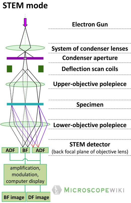
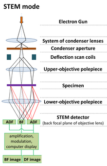
Transmission Electron Microscopy | MRSEC The Electron Microscopy Facility is a joint BSD/PSD resource available to all campus researchers. Users have access to an FEI Tecnai F30 scanning/transmission electron microscope. The microscope has a point-to-point resolution of 0.2 nm when operated in the TEM mode and a spatial resolution of 0.2 nm for the STEM mode.The facility is located in ... A Guide to Scanning Transmission Electron Microscopy (STEM) Cryogenic electron microscopy (cryo-EM), a later variation of the electron microscope, also led to the 2017 Nobel Prize in chemistry. Continue reading: Using ZnO Nanowires for 4D STEM Technology References and Further Reading. Liu J. (2005) Scanning transmission electron microscopy and its application to the study of nanoparticles and ...
Transmission electron microscopy DNA sequencing - Wikipedia Transmission electron microscopy (TEM) produces high magnification, high resolution images by passing a beam of electrons through a very thin sample. Whereas atomic resolution has been demonstrated with conventional TEM, further improvement in spatial resolution requires correcting the spherical and chromatic aberrations of the microscope lenses.
Transmission electron micrograph labeled
Transmission electron micrograph showing immunogold labeled DipA in the ... Download scientific diagram | Transmission electron micrograph showing immunogold labeled DipA in the outer membrane of the B. burgdorferi Ospless mutant B313. Ultrathin cryosections were prepared ... Animal Cell Electron Microscope Labelled - Q14 Draw a large diagram of ... This is because of the way that the cell was sectioned (cut) before it was viewed on the transmission electron microscope. Source: hi-static.z-dn.net. Here is an electron micrograph of an animal cell with the labels superimposed: Animal cell electron micrograph labelling. The cell membrane is what controls the entry and exit of any substances ... Label This Transmission Electron Micrograph / Microscopy Innovations ... Label the transmission electron micrograph of the nucleus. Transmission electron micrographs of hela cell sections labeled in . Label the transmission electron micrograph of the nucleus. Fluorescence microscopy in combination with tem and an ion beam analysis (iba, which allows the evaluation of the chemical elemental distribution) has allowed .
Transmission electron micrograph labeled. Solved Label the transmission electron micrograph based on - Chegg Expert Answer nucleus is the house of the genetic material which contains all the h … View the full answer Transcribed image text: Label the transmission electron micrograph based on the hints provided Mitochondrion Heterochromatin Plasma cell Nucleus Rough endoplasmic reticulum Nucleolus Previous question Next question Transmission Electron Microscopy - Penn State College of Medicine Research The JEOL 1400 TEM (Room C1727) is capable of generating ultra-structural nanoscale images from fixed cell/tissue samples or multiplexed immune-labeled samples. Computer-controlled operations Resolution up to 3 Angstroms Magnification up to 370,000X Capable of collecting data suitable for 3D reconstructions of negative-stained samples Transmission Electron Microscope (TEM)- Definition, Principle, Images Parts of Transmission Electron Microscope (TEM) Their working mechanism is enabled by the high-resolution power they produce which allows it to be used in a wide variety of fields. It has three working parts which include: Electron gun Image producing system Image recording system Electron gun Chloroplast Electron Microscope - transmission electron micrograph of ... Chloroplast Electron Microscope - 18 images - chloroplast structure under microscope micropedia, cell biology chloroplast, light and electron microscope images ccber, labled diagram of a chloroplast micrograph photo by akucic biology,
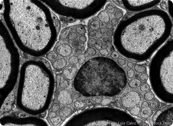
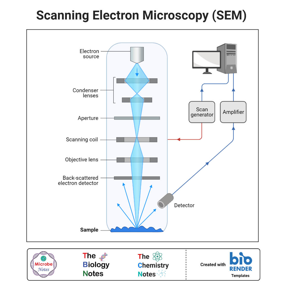
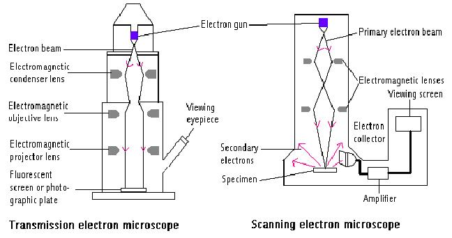
Electron Microscope- Definition, Principle, Types, Uses, Labeled Diagram There are two types of electron microscopes, with different operating styles: 1. Transmission Electron Microscope (TEM) The transmission electron microscope is used to view thin specimens through which electrons can pass generating a projection image. The TEM is analogous in many ways to the conventional (compound) light microscope. Transmission electron microscopy - Wikipedia Transmission electron microscopy ( TEM) is a microscopy technique in which a beam of electrons is transmitted through a specimen to form an image. The specimen is most often an ultrathin section less than 100 nm thick or a suspension on a grid. Transmission Electron Micrograph of transfected HL-1 cells labeled for ... Transmission Electron Micrograph of transfected HL-1 cells labeled for TMEM43 with immunogold. A and B. Single immunogold labeling experiments used 15 nm gold particles to label GFP. A.... Transmission Electron Microscope (With Diagram) - Biology Discussion The final image in a TEM is known as transmission electron micrograph. The salts of some heavy metals, e.g., lead; osmium, tungsten and uranium are often used for staining. These heavy metal stains are used to increase the contrast between ultra structures and the background.
Transmission Electron Micrographs of Negatively Stained Salmonella ... This low magnification micrograph depicts a single bacterium possessing multiple flagella; the arrow indicates one of these flagella. Protein aggregates from the media are visible as light-colored circles. Bar = 1 µm. Figure 2: Negatively Stained Transmission Electron Micrograph of Salmonella typhimurium (Labeled view). Label This Transmission Electron Micrograph - Kaiden Brown Label this transmission electron micrograph of relaxed sarcomeres by clicking and dragging the labels to the correct location . Transmission electron microscopy (tem) is one of the oldest technologies and still. Molecular labeling for correlative microscopy: Fluorescence microscopy in combination with tem and an ion beam analysis (iba, which ... Label This Transmission Electron Micrograph Of A Relaxed ... - Blogger Label this transmission electron micrograph of relaxed sarcomeres by clicking and dragging the labels to the correct location . Label the following image using the terms provided. Note how the sarcomeres are extended to only approximately 120 % . IMG_2132 - FIGURES Label this transmission electron from Electron Micrographs - University of Oklahoma Health Sciences Center Electron Micrographs Below is a collection of electron micrographs with labelled subcellular structures that you should be able to identify. Also, be sure to observe any electron micrographs which are made available in the laboratory by the instructor.
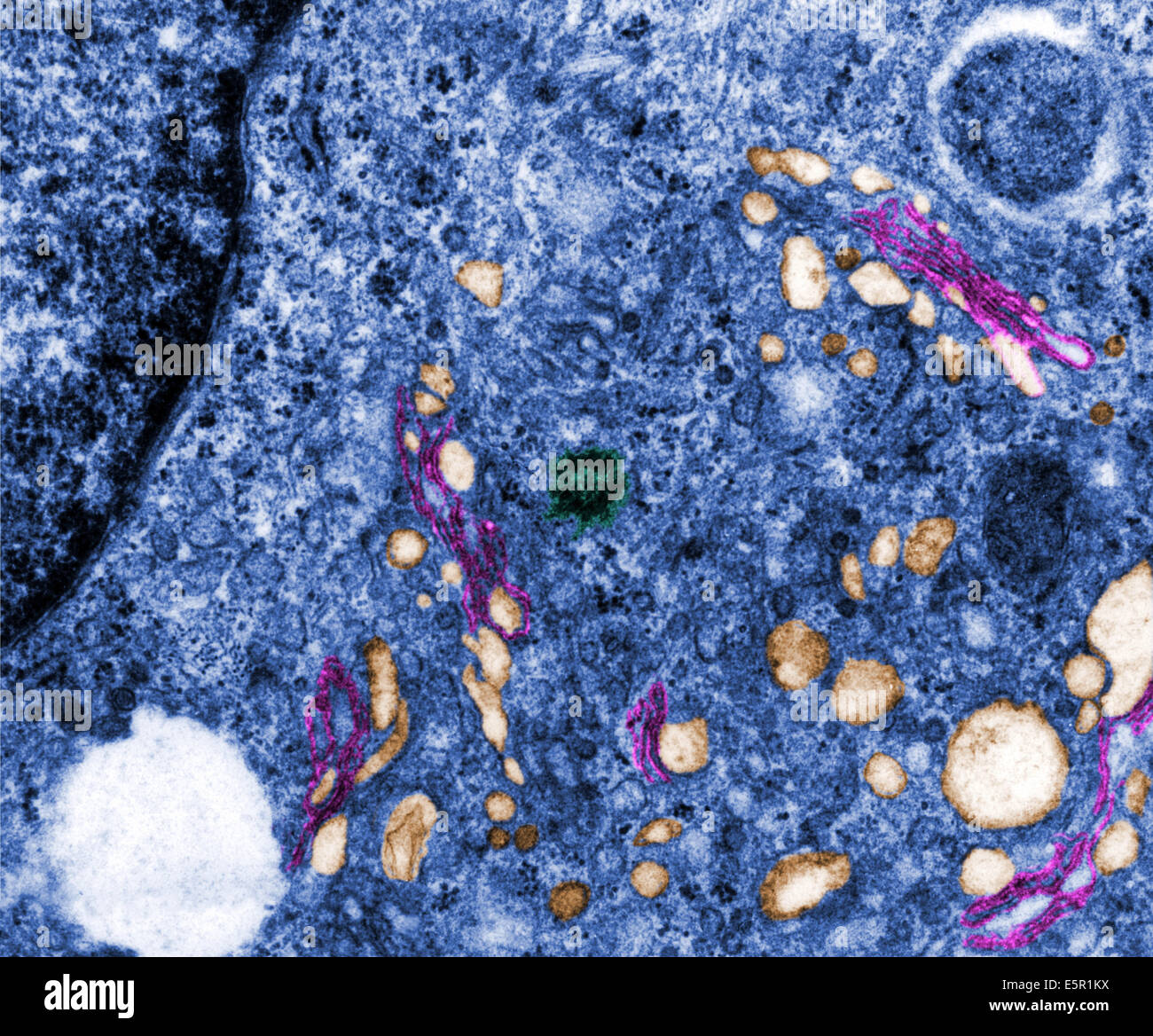
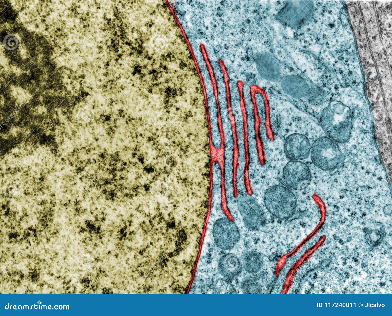
Transmission electron micrograph (TEM) of the Golgi apparatus ...
Transmission electron microscopy DNA sequencing - dnareplicationsystem Transmission electron microscopy DNA sequencing is a single-molecule sequencing technology that uses transmission electron microscopy techniques.The method was conceived and developed in the 1960s and 70s, but lost favor when the extent of damage to the sample was recognized. DNA is visible under the electron microscope; however, it must be labeled with heavy atoms so that the DNA bases can be ...
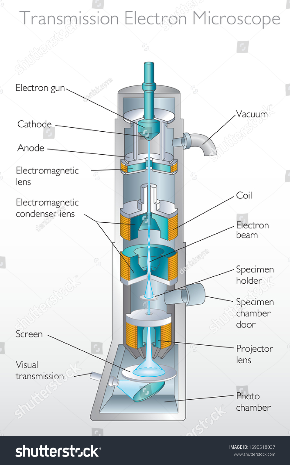
Diagram Transmission Electron Microscope Quantum Physics ...
Solved Label this transmission electron micrograph of - Chegg Anatomy and Physiology questions and answers Label this transmission electron micrograph of relaxed sarcomeres by clicking and dragging the labels to the correct location Sarcamere 1 band (light) Z disc Mline Aband (dark) H zone

A Technical Introduction to Transmission Electron Microscopy ...
Transmission electron microscopy characterization of fluorescently ... Results: Using transmission electron microscopy (TEM), we verify that N-terminal labeling of Aβ40 with AMCA, TAMRA, and Hilyte-Fluor 488 tags does not prevent the formation of protofibrils and amyloid fibrils of various widths. We also measure the two-photon action cross-section of Aβ40 labelled with Hilyte Fluor 488 and demonstrate that this ...

Transmission electron microscopy images of immunogold-labeled ...
Scanning & Transmission Electron Microscopy Reveals Graphene ... February 5th, 2021 - Updated October 1st, 2021 & March 12th, 2022! Author: Robert O Young CPC, MSc, DSc, PhD, Naturopathic Practitioner 16th Revision Phase Contrast, Dark Field, Bright Field Microscopy, Transmission and Scanning Electron Microscopy and Energy-Dispersive X-ray Spectroscopy Reveal the Poisonous Ingredients in the CoV-19 Vaccines!
Electron Microscope Principle, Uses, Types and Images ...
PPTX Transmission Electron Micrographs of Negatively Stained Salmonella ... Negatively Stained Transmission Electron Micrograph of Salmonella typhimurium S. typhimurium negatively stained with 1% uranyl acetate. This low magnification micrograph depicts a single bacterium possessing multiple flagella; the arrow indicates one of these flagella. Protein aggregates from the media are visible as light-colored circles. Bar ...

The Transmission Electron Microscope | CCBER
Transmission Electron Microscopy - an overview | ScienceDirect Topics Transmission Electron Microscopy. Transmission electron microscopy (TEM) is a powerful tool for examination of the microanatomy of biological tissues, cells and organisms including nematodes. ... Clusters of wheat germ agglutinin-labeled gold granules were located all over the surface of milk fat globules using SEM, suggesting the presence of N ...

Transmission electron microscopy - Wikipedia
Microscope Types (with labeled diagrams) and Functions The shorter wavelength of electrons compared to visible light photons helps the observer achieve a very high resolving power compared to normal microscopes thereby aiding observers to see very tiny objects clearly. Electron microscope labeled diagram The different types of electron microscopes are: Transmission Electron Microscope

Transmission electron microscopy - Wikipedia
PDF TRANSMISSION ELECTRON MICROSCOPY - University of Florida transmission electron microscopy negative stains, immuno-specific labeling (isem), thin sectioning of fixed and embedded material pm 7/126 (1) electron microscopy in diagnosis of plant viruses • eppo bulletin (2015) 45:450-453 - electron microscopy can be used for detection or identificatio n of viruses in tissue extracts of infected plant ...
Nuclear envelope. TEM stock image. Image of micrograph ...
Transmission Electron Microscopy (TEM) - Warwick The transmission electron microscope is a very powerful tool for material science. A high energy beam of electrons is shone through a very thin sample, and the interactions between the electrons and the atoms can be used to observe features such as the crystal structure and features in the structure like dislocations and grain boundaries.

Labeling the Cell Flashcards | Quizlet
Looking at the Structure of Cells in the Microscope Determining the detailed structure of the membranes and organelles in cells requires the higher resolution attainable in a transmission electron microscope. Specific macromolecules can be localized with colloidal gold linked to antibodies. Three-dimensional views of the surfaces of cells and tissues are obtained by scanning electron microscopy.

Scanning transmission electron microscopy - Wikipedia
Labeling the Cell Flashcards | Quizlet Label the transmission electron micrograph of the nucleus. membrane bound organelles golgi apparatus, mitochondrion, lysosome, peroxisome, rough endoplasmic reticulum nonmembrane bound organelles ribosomes, centrosome, proteasomes cytoskeleton includes microfilaments, intermediate filaments, microtubules Identify the highlighted structures
Transmission Electron Microscope: Definition, Parts, Working ...
Label This Transmission Electron Micrograph / Microscopy Innovations ... Label the transmission electron micrograph of the nucleus. Transmission electron micrographs of hela cell sections labeled in . Label the transmission electron micrograph of the nucleus. Fluorescence microscopy in combination with tem and an ion beam analysis (iba, which allows the evaluation of the chemical elemental distribution) has allowed .

Solved Label the transmission electron micrograph based on ...
Animal Cell Electron Microscope Labelled - Q14 Draw a large diagram of ... This is because of the way that the cell was sectioned (cut) before it was viewed on the transmission electron microscope. Source: hi-static.z-dn.net. Here is an electron micrograph of an animal cell with the labels superimposed: Animal cell electron micrograph labelling. The cell membrane is what controls the entry and exit of any substances ...
Electron Microscope Principle, Uses, Types and Images ...
Transmission electron micrograph showing immunogold labeled DipA in the ... Download scientific diagram | Transmission electron micrograph showing immunogold labeled DipA in the outer membrane of the B. burgdorferi Ospless mutant B313. Ultrathin cryosections were prepared ...

Solved Mitochondrion Nucleus Vesicle Peroxisome | Chegg.com
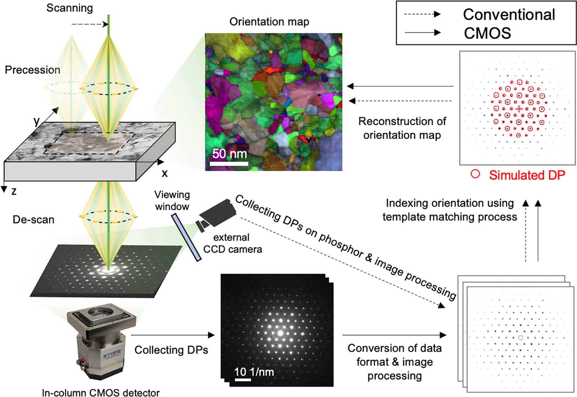
Automated Crystal Orientation Mapping by Precession Electron ...

Transmission Electron Micrographs of Plasmodesmata at Cell ...

Transmission electron microscope for USPIO-labeled cells ...

Transmission electron micrograph of turkey spermatozoa ...

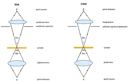
Ray diagrams showing the important optical elements for (a ...
A tour of the cell: View as single page

Transmission Electron Microscopy - an overview ...
What is Transmission Electron Microscopy?
Transmission electron microscopy - Wikipedia

Transmission electron microscopy - Wikipedia
Transmission Electron Microscope (TEM)- Definition, Principle ...
Transmission Electron Microscope: Definition, Parts, Working ...
Transmission Electron Microscopy | Central Microscopy ...

What is Transmission Electron Microscopy?
Electron Microscope- Definition, Principle, Types, Uses ...
Transmission Electron Microscope (TEM)- Definition, Principle ...
Scanning transmission electron microscopy - Wikipedia
Farlander Central

8.2: Transmission Electron Microscopy - Chemistry LibreTexts

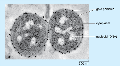
Transmission electron microscopy (TEM). Ten-nanometre gold ...

Transmission electron micrographs of HeLa cell sections ...

Types of Microscopes - Exploring MICROSCOPY

Transmission electron microscopy (TEM) of graphene ...
Post a Comment for "38 transmission electron micrograph labeled"