38 diagram of a microscope with labels
Wikipedia:Citation needed - Wikipedia To ensure that all Wikipedia content is verifiable, Wikipedia provides a means for anyone to question an uncited claim.If your work has been tagged, please provide a reliable source for the statement, and discuss if needed.. You can add a citation by selecting from the drop-down menu at the top of the editing box.In markup, you can add a citation manually using ref tags. PDF Parts of a Microscope Printables - Homeschool Creations Label the parts of the microscope. You can use the word bank below to fill in the blanks or cut and paste the words at the bottom. Microscope Created by Jolanthe @ HomeschoolCreations.net. Parts of a eyepiece arm stageclips nosepiece focusing knobs illuminator stage objective lenses
quizlet.com › 515111566 › ch-8-mastering-biologych 8 mastering biology Flashcards | Quizlet Drag the labels onto the diagram to identify the stages of the cell cycle. 1. most of the cells life is spent in interphase 2. in phosphase microtubules form the mitototic spindle
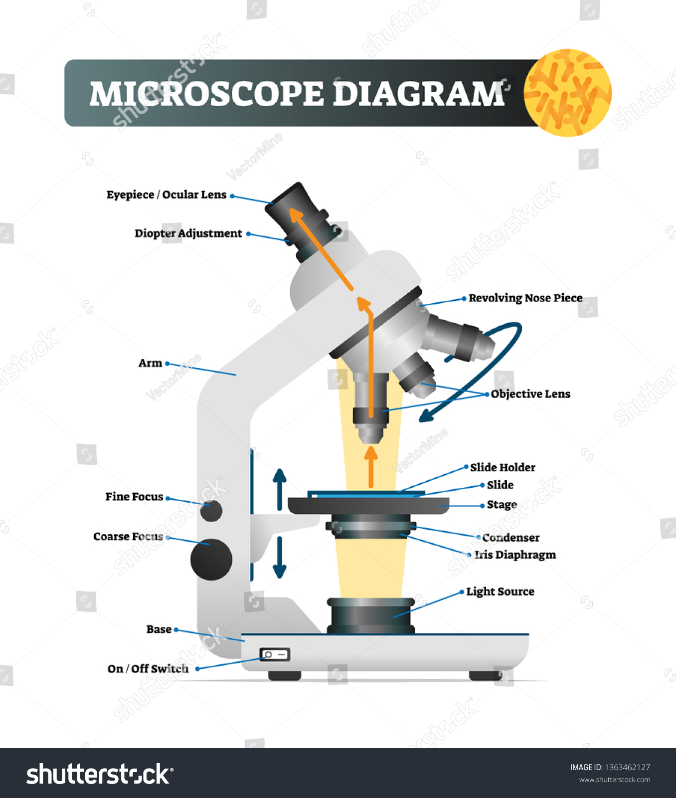
Diagram of a microscope with labels
Comparative genomic hybridization - Wikipedia Comparative genomic hybridization (CGH) is a molecular cytogenetic method for analysing copy number variations (CNVs) relative to ploidy level in the DNA of a test sample compared to a reference sample, without the need for culturing cells. The aim of this technique is to quickly and efficiently compare two genomic DNA samples arising from two sources, which are most often … Microscope Types (with labeled diagrams) and Functions Simple microscope labeled diagram Simple microscope functions It is used in industrial applications like: Watchmakers to assemble watches Cloth industry to count the number of threads or fibers in a cloth Jewelers to examine the finer parts of jewelry Miniature artists to examine and build their work Also used to inspect finer details on products mastering ch.13 Flashcards | Quizlet Complete the diagram to show the life cycle of a typical animal. Follow these steps: 1. First, drag blue labels onto blue targets only to identify each stage of the life cycle. 2. Next, drag pink labels onto pink targets only to identify the process by which each stage occurs. 3.
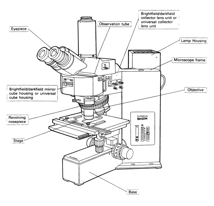
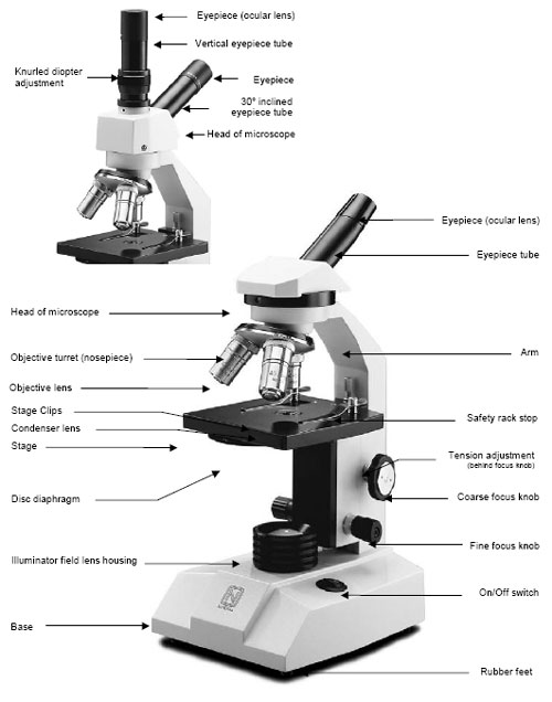
Diagram of a microscope with labels. Simple Microscope - Diagram (Parts labelled), Principle, Formula and Uses A simple microscope consists of Optical parts Mechanical parts Labeled Diagram of simple microscope parts Optical parts The optical parts of a simple microscope include Lens Mirror Eyepiece Lens A simple microscope uses biconvex lens to magnify the image of a specimen under focus. A Study of the Microscope and its Functions With a Labeled Diagram ... These labeled microscope diagrams and the functions of its various parts, attempt to simplify the microscope for you. However, as the saying goes, 'practice makes perfect', here is a blank compound microscope diagram and blank electron microscope diagram to label. Download the diagrams and practice labeling the different parts of these ... › products › microscopeMicroscope Objective Lens | Products | Leica Microsystems The objective lens is a critical part of the microscope optics. The microscope objective is positioned near the sample, specimen, or object being observed. It has a very important role in imaging, as it forms the first magnified image of the sample. The numerical aperture (NA) of the objective indicates its ability to gather light and largely determines the microscope’s resolution, the ... Binocular Microscope Anatomy - Parts and Functions with a Labeled Diagram The nose piece of a microscope, Head part of the microscope, Ocular lens or eyepiece of the microscope, Diopter adjustment of the eyepiece All of these parts are identified in a light microscope labeled diagram. So, first, make sure you can identify all these parts from this labeled diagram. Parts of the compound microscope
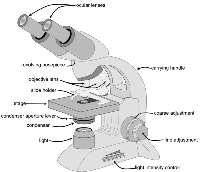
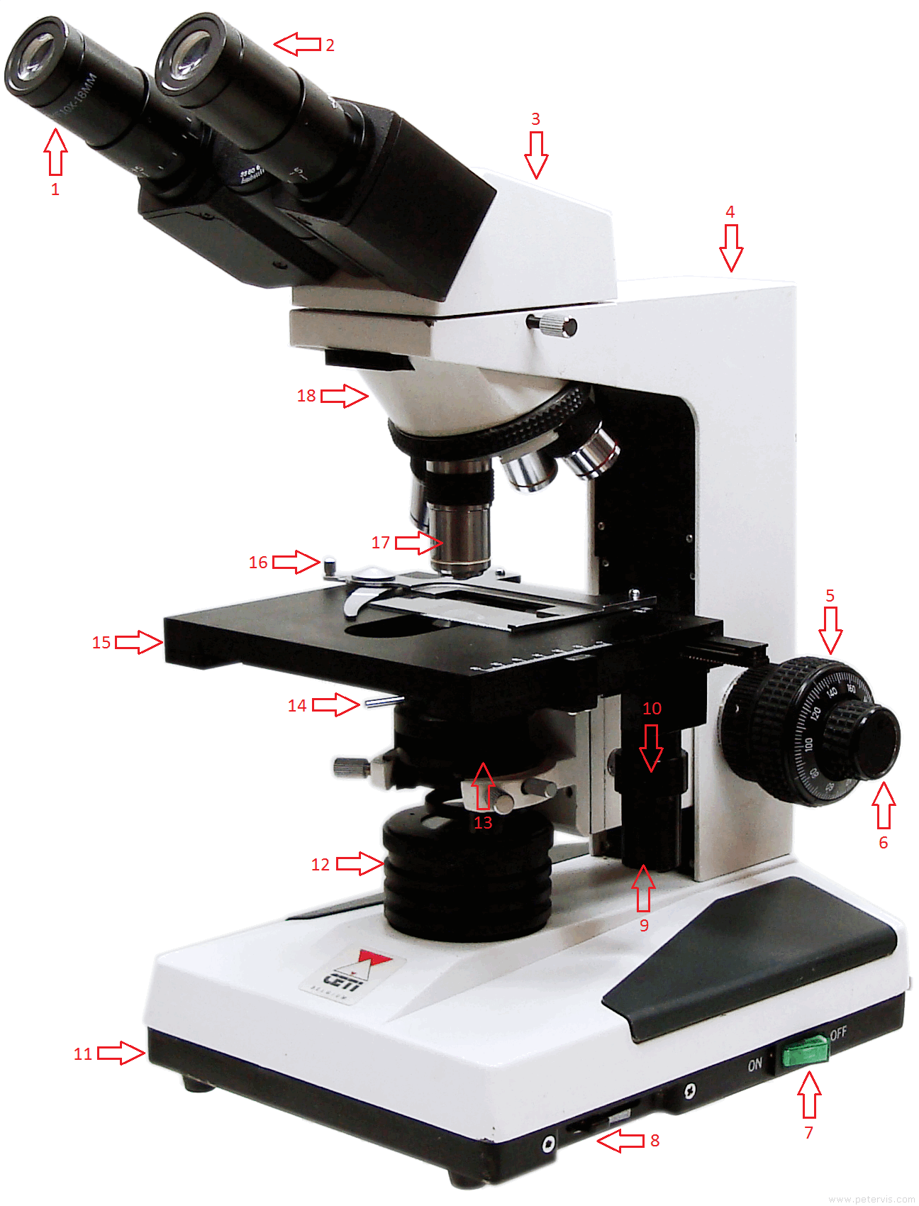
Achiever Papers - We help students improve their academic standing Professional academic writers. Our global writing staff includes experienced ENL & ESL academic writers in a variety of disciplines. This lets us find the most appropriate writer for any type of assignment. Compound Microscope - Diagram (Parts labelled), Principle and Uses See: Labeled Diagram showing differences between compound and simple microscope parts Structural Components The three structural components include 1. Head This is the upper part of the microscope that houses the optical parts 2. Arm This part connects the head with the base and provides stability to the microscope. Parts of a microscope with functions and labeled diagram - Microbe Notes Q. List down the 18 parts of a Microscope. 1. Ocular Lens (Eye Piece) 2. Diopter Adjustment 3. Head 4. Nose Piece 5. Objective Lens 6. Arm (Carrying Handle) 7. Mechanical Stage 8. Stage Clip 9. Aperture 10. Diaphragm 11. Condenser 12. Coarse Adjustment 13. Fine Adjustment 14. Illuminator (Light Source) 15. Stage Controls 16. Base 17. Assignment Essays - Best Custom Writing Services Get 24⁄7 customer support help when you place a homework help service order with us. We will guide you on how to place your essay help, proofreading and editing your draft – fixing the grammar, spelling, or formatting of your paper easily and cheaply.
Microscope Parts, Function, & Labeled Diagram - slidingmotion Microscope parts labeled diagram gives us all the information about its parts and their position in the microscope. Microscope Parts Labeled Diagram The principle of the Microscope gives you an exact reason to use it. It works on the 3 principles. Magnification Resolving Power Numerical Aperture. Parts of Microscope Head Base Arm Eyepiece Lens The Parts of a Microscope (Labeled) Printable - TeacherVision The Parts of a Microscope (Labeled) Printable. Download. Add to Favorites. Share. This diagram labels and explains the function of each part of a microscope. Use this printable as a handout or transparency to help prepare students for working with laboratory equipment. Grade: Parts of Stereo Microscope (Dissecting microscope) – labeled diagram ... Labeled part diagram of a stereo microscope Major structural parts of a stereo microscope. ... (based on color bands and their respective labels), the objectives of a dissecting microscope are located in a cylindrical cone and, therefore, are not directly seen. For the stereo microscope that comes with multiple objective lens sets (fixed power ... Microscope, Microscope Parts, Labeled Diagram, and Functions The description given below summarize the brief description of microscope parts used to visualize the microscopic specimens such as animal cells, plant cells, microbes, bacteria, viruses, microorganisms etc. The Microscopes parts divided into three different structural parts Head, Base, and Arms.
4,800 Microscope labeled Images, Stock Photos & Vectors ...
› 6-label-the-microscopeLabel the microscope — Science Learning Hub Jun 08, 2018 · All microscopes share features in common. In this interactive, you can label the different parts of a microscope. Use this with the Microscope parts activity to help students identify and label the main parts of a microscope and then describe their functions. Drag and drop the text labels onto the microscope diagram. If you want to redo an ...

Microscope With Labels clip art | Microscope parts ...
UV Properties of Plastics: Transmission and Resistance - Cole-Parmer May 03, 2021 · One of the main problems of considering the effect of UV rays on polymers is the intensity related to: stratospheric ozone, clouds, altitude, the position of the sun height (time of day and time of year), and reflection. The complexity of the effects can be seen in a global plot of UV levels dark green being the highest:

Compound Microscope Parts, Functions, and Labeled Diagram ...
Labeled Microscope Diagram Clipart Free Download 291 Labeled Microscope Diagram clipart free images in AI, SVG, EPS or CDR. Realistic brand cosmetic bottles. Minimalist labeled black containers design, beauty products packages, pumps and spray mockups. Vector set.

File:Labelledmicroscope.gif - Wikibooks, open books for an ...
Microscope Objective Lens | Products | Leica Microsystems The objective lens is a critical part of the microscope optics. The microscope objective is positioned near the sample, specimen, or object being observed. It has a very important role in imaging, as it forms the first magnified image of the sample. The numerical aperture (NA) of the objective indicates its ability to gather light and largely determines the microscope’s …

Parts of the Microscope with Labeling (also Free Printouts ...
Microscope Parts and Functions Most specimens are mounted on slides, flat rectangles of thin glass. The specimen is placed on the glass and a cover slip is placed over the specimen. This allows the slide to be easily inserted or removed from the microscope. It also allows the specimen to be labeled, transported, and stored without damage.

5 Important Types of Microscopes used in Biology (With Diagram)
Compound Microscope: Definition, Diagram, Parts, Uses, Working ... - BYJUS Compound microscope is a type of optical microscope that is used for obtaining a high-resolution image. There are more than two lenses in a compound microscope. Learn about the working principle, parts and uses of a compound microscope along with a labeled diagram here.

Dissecting Stereo Microscope Parts and Functions
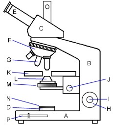
Microscope Diagram - Label Diagram | Quizlet The bottom of the microscope, used for support. ocular lens. Eyepiece of a microscope. Diaphragm. Regulates the amount of light on the specimen. nosepiece of microscope. holds the objective lenses. objective lens. The lens on a light microscope that is closest to the stage.

microscope drawing with label - Clip Art Library
Microscope, Microscope Parts, Labeled Diagram, and Functions (2022) The description given below summarize the brief description of microscope parts used to visualize the microscopic specimens such as animal cells, plant cells, microbes, bacteria, viruses, microorganisms etc. The Microscopes parts divided into three different structural parts Head, Base, and Arms.

Microscope labeling
Compound Microscope Parts, Functions, and Labeled Diagram Compound Microscope Definitions for Labels. Eyepiece (ocular lens) with or without Pointer: The part that is looked through at the top of the compound microscope. Eyepieces typically have a magnification between 5x & 30x. Monocular or Binocular Head: Structural support that holds & connects the eyepieces to the objective lenses.

Label the Microscope Diagram | Download Scientific Diagram
› books › NBK26880Looking at the Structure of Cells in the Microscope ... Many light-microscope techniques are available for observing cells. Cells that have been fixed and stained can be studied in a conventional light microscope, while antibodies coupled to fluorescent dyes can be used to locate specific molecules in cells in a fluorescence microscope. Living cells can be seen with phase-contrast, differential ...
Simple Microscope - Definition, Diagram, FAQs
Label the microscope — Science Learning Hub Jun 08, 2018 · All microscopes share features in common. In this interactive, you can label the different parts of a microscope. Use this with the Microscope parts activity to help students identify and label the main parts of a microscope and then describe their functions.. Drag and drop the text labels onto the microscope diagram. If you want to redo an answer, click on the …
Jual PREORDER Microscope Set 100X-1200X Children Kids ...
rsscience.com › stereo-microscopeParts of Stereo Microscope (Dissecting microscope) – labeled ... Labeled part diagram of a stereo microscope Major structural parts of a stereo microscope. There are three major structural parts of a stereo microscope. The viewing Head includes the upper part of the microscope, which houses the most critical optical components, including the eyepiece, objective lens, and light source of the microscope.

Parts of a microscope with functions and labeled diagram
Compound Microscope Parts, Functions, and Labeled Diagram Nov 18, 2020 · Parts of a Compound Microscope Each part of thenbsp compound microscope serves its own unique function, with each being important to the function of the scope as a whole. The individual parts of a compound microscope can vary heavily depending on the configuration & applications that the scope is being used for. Common compound microscope parts include: …

Draw a well labelled diagram of a microscope. - Brainly.in
Simple Microscope - Parts, Functions, Diagram and Labelling Parts of the optical parts are as follows: Mirror - A simple microscope has a plano-convex mirror and its primary function is to focus the surrounding light on the object being examined. Lens - The biconvex lens is placed above the stage and its function is to magnify the size of the object being examined.
Jual PREORDER 1600X Student Biological Microscope 2MP USB ...
Compound Microscope Parts - Labeled Diagram and their Functions Labeled diagram of a compound microscope Major structural parts of a compound microscope There are three major structural parts of a compound microscope. The head includes the upper part of the microscope, which houses the most critical optical components, and the eyepiece tube of the microscope.
MICROBIO 16 Parts of a Compound Microscope with Diagram and ...
Microscope labeled diagram - SlideShare Follow 1. The Microscope Image courtesy of: Microscopehelp.com Basic rules to using the microscope 1. You should always carry a microscope with two hands, one on the arm and the other under the base. 2. You should always start on the lowest power objective lens and should always leave the microscope on the low power lens when you finish using it.

Label Microscope Diagram | Microscope parts, Microscope ...
› science › articleCompatibility studies between N-A-S-H and C-A-S-H gels. Study ... Sep 01, 2011 · Finally TEM characterization was conducted using a JEOL 200EX. TEM microscope fitted with a LINK AN10/855 EDX analyzer. 2.2. Direct mixing of gels. Two series of 50/50 wt.% mixtures of dried pre-formed C-S-H and N-A-S-H and C-A-S-H and N-A-S-H gels were re-dispersed in ultra pure degassed water with continuous stirring. Solid samples were taken ...

Simple Microscope - Parts, Functions, Diagram and Labelling ...
en.wikipedia.org › wiki › Electron_microscopeElectron microscope - Wikipedia An electron microscope is a microscope that uses a beam of accelerated electrons as a source of illumination. As the wavelength of an electron can be up to 100,000 times shorter than that of visible light photons , electron microscopes have a higher resolving power than light microscopes and can reveal the structure of smaller objects.

Biology Notes for A level: #3. Microscopy
mastering ch.13 Flashcards | Quizlet Complete the diagram to show the life cycle of a typical animal. Follow these steps: 1. First, drag blue labels onto blue targets only to identify each stage of the life cycle. 2. Next, drag pink labels onto pink targets only to identify the process by which each stage occurs. 3.

Simple Microscope - Diagram (Parts labelled), Principle ...
Microscope Types (with labeled diagrams) and Functions Simple microscope labeled diagram Simple microscope functions It is used in industrial applications like: Watchmakers to assemble watches Cloth industry to count the number of threads or fibers in a cloth Jewelers to examine the finer parts of jewelry Miniature artists to examine and build their work Also used to inspect finer details on products

Label the diagram of the microscope and explain the role of ...
Comparative genomic hybridization - Wikipedia Comparative genomic hybridization (CGH) is a molecular cytogenetic method for analysing copy number variations (CNVs) relative to ploidy level in the DNA of a test sample compared to a reference sample, without the need for culturing cells. The aim of this technique is to quickly and efficiently compare two genomic DNA samples arising from two sources, which are most often …

Draw a labelled diagram of a compound microscope.

Compound Microscope: Parts of Compound Microscope

Schematic diagram of the of the WFLPCF microscope. | Download ...
Glossary of terms used in microscopy – Quekett Microscopical Club

Labelling a Microscope Diagram | Quizlet
Labelled Diagram of Microscope Parts
INDIAN INSTITUTE OF TECHNOLOGY
Microscope Maintenance Tips

How to draw Microscope diagram for beginners - step by step

Labeled Microscope Diagram | Microscope parts, Science fair ...

The Microscope
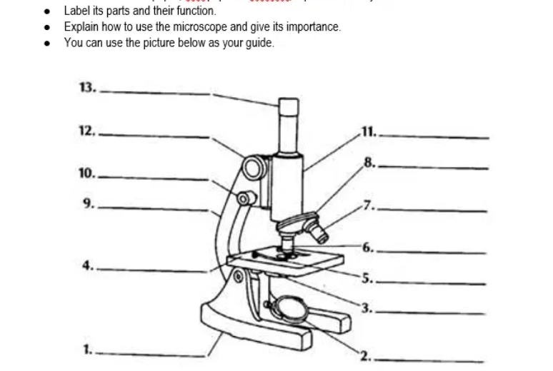
Answered: Label its parts and their function.… | bartleby

Ecology Test Day Microscope Introduction Day (Test day ...

Lab 1: The Laboratory Microscope

Lable the microscope worksheet
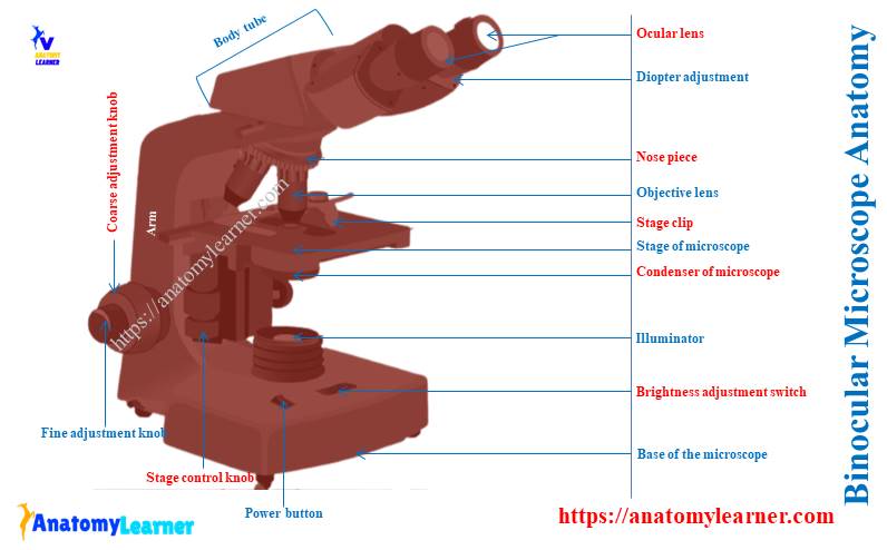
Binocular Microscope Anatomy - Parts and Functions with a ...

Microscope, Microscope Parts, Labeled Diagram, and Functions
Post a Comment for "38 diagram of a microscope with labels"